
Professional medical service of Obstetrical Ultrasound
What is Obstetrical Ultrasound?
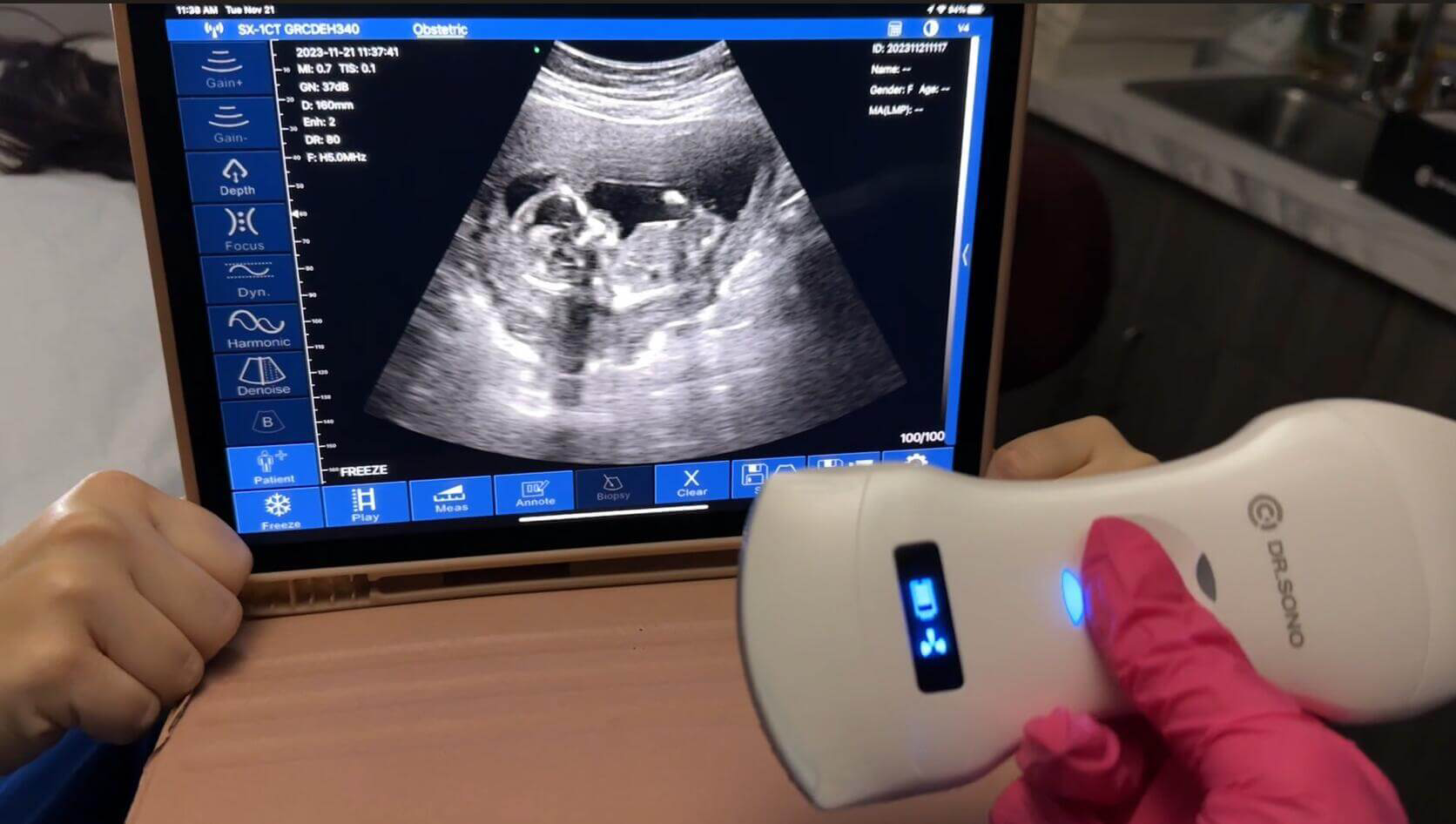
Obstetrical ultrasound is a noninvasive medical imaging test that uses sound waves to create real-time images of the embryo or fetus in a woman’s uterus, as well as the uterus and ovaries of the mother. It uses a small probe and gel applied directly to the skin to transmit high-frequency sound waves, which a computer processes into detailed images. Unlike x-rays, ultrasound does not use radiation, making it a safe and painless diagnostic tool.
In certain cases, blood flow in the umbilical cord, fetus, or placenta may also be evaluated using this method.
Why Should I Do It?
Obstetrical ultrasound is a key clinical tool used to:
- Establish the presence of a living embryo or fetus.
- Estimate the age of the pregnancy.
- Diagnose congenital abnormalities of the fetus.
- Evaluate the position of the fetus.
- Assess the position of the placenta.
- Determine if there are multiple pregnancies.
- Measure the amount of amniotic fluid surrounding the baby.
- Check for any opening or shortening of the cervix.
- Monitor fetal growth and assess fetal well-being.
Some doctors may also use 3-D ultrasound to further assess fetal development.
Preparation Guidelines
- Clothing:
- Wear a loose-fitting, two-piece outfit to the exam. Only your lower abdominal area will need to be exposed.
- Specialized Techniques:
- For early pregnancies or to closely evaluate the cervix, a transvaginal ultrasound may be performed. This involves inserting a small transducer into the vagina for more detailed imaging.